


Peptide substrates have emerged as game - changers in the field of tissue engineering. As a supplier of high - quality peptide substrates, I've seen firsthand how these tiny molecules can have a huge impact on cell adhesion and proliferation. In this blog, I'll break down how peptide substrates work their magic in tissue engineering.
The Basics of Cell Adhesion and Proliferation in Tissue Engineering
Before we dive into the role of peptide substrates, let's quickly go over what cell adhesion and proliferation mean in the context of tissue engineering. Tissue engineering aims to create functional tissues by combining cells, scaffolds, and signaling molecules. Cell adhesion is the process by which cells attach to each other or to a surface. It's like the foundation of a building; without proper adhesion, cells can't form organized structures. Proliferation, on the other hand, is all about cell growth and division. Once cells are attached, they need to multiply to form enough tissue to be useful.
How Peptide Substrates Promote Cell Adhesion
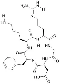
Peptide substrates act as molecular glue for cells. They contain specific amino acid sequences that can bind to cell surface receptors. One of the most well - known sequences is the arginine - glycine - aspartic acid (RGD) sequence. This sequence is recognized by integrins, which are cell surface receptors involved in cell adhesion. When a peptide substrate with the RGD sequence is present, cells can easily attach to it.
Think of it like a lock - and - key mechanism. The integrins on the cell surface are the locks, and the RGD sequence on the peptide substrate is the key. When the key fits into the lock, the cell can firmly attach to the substrate. This attachment not only provides physical support for the cell but also triggers a series of intracellular signaling pathways. These pathways can regulate gene expression, cell survival, and migration, all of which are crucial for tissue development.


For example, in a tissue engineering scaffold, peptide substrates can be incorporated into the material. This makes the scaffold more cell - friendly, allowing cells to adhere more efficiently. It's like making a welcome mat for the cells, encouraging them to settle down and start building the tissue.
Impact on Cell Proliferation
Peptide substrates don't just stop at promoting cell adhesion; they also play a vital role in cell proliferation. Once cells are attached to the peptide substrate, they receive signals that tell them to start dividing. These signals can be in the form of growth factors or other signaling molecules that are either bound to the peptide substrate or released in response to cell - substrate interaction.
Some peptide substrates can mimic the extracellular matrix (ECM), which is the natural environment where cells live in the body. The ECM provides a rich source of growth factors and other bioactive molecules. By mimicking the ECM, peptide substrates can create a similar microenvironment that supports cell growth.
For instance, certain peptide substrates can bind to growth factors and present them to the cells in a more effective way. This enhances the cells' ability to respond to these growth factors, leading to increased proliferation. It's like giving the cells a boost of energy to keep multiplying.
Examples of Peptide Substrates in Action
Let's take a look at some specific peptide substrates that are commonly used in tissue engineering.
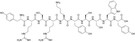
Calpain Inhibitor VI CAS 190274 - 53 - 4 is a peptide substrate that has shown potential in promoting cell adhesion and proliferation. It can inhibit the activity of calpain, an enzyme that is involved in cell death and migration. By inhibiting calpain, this peptide substrate can help cells stay alive and continue to divide.
Suc - IIW - AMC is another interesting peptide substrate. It can interact with cell surface receptors and trigger intracellular signaling pathways that are beneficial for cell growth. It has been used in various tissue engineering applications, especially in the development of artificial skin and cartilage.
Z - LLY - FMK CAS 133410 - 84 - 1 is a peptide substrate that can inhibit caspase activity. Caspases are enzymes involved in apoptosis, or programmed cell death. By inhibiting caspases, this peptide substrate can prevent cell death and promote cell survival and proliferation.
Advantages of Using Our Peptide Substrates
As a supplier, we take pride in offering high - quality peptide substrates. Our products are carefully synthesized and purified to ensure consistent quality. We also offer a wide range of peptide substrates with different sequences and functions, so you can choose the ones that best suit your tissue engineering needs.
Our peptide substrates are cost - effective, which is important for researchers and companies working on tissue engineering projects. We understand that budget is often a concern, and we strive to provide the best value for your money.
In addition, we have a team of experts who can provide technical support. Whether you have questions about the properties of a specific peptide substrate or need advice on how to use it in your experiments, we're here to help.
Conclusion and Call to Action
Peptide substrates are powerful tools in tissue engineering, promoting both cell adhesion and proliferation. They offer a way to create more effective tissue engineering scaffolds and improve the success rate of tissue regeneration.
If you're involved in tissue engineering research or development, I encourage you to explore our range of peptide substrates. We're confident that our products can make a difference in your projects. Whether you're working on a small - scale research experiment or a large - scale tissue engineering production, we have the right peptide substrates for you.
To learn more about our peptide substrates or to start a procurement discussion, feel free to reach out. We're looking forward to working with you to advance the field of tissue engineering.
References
- Hubbell JA. Biomaterials in tissue engineering. BioMaterials. 1995;16(12):1079 - 1092.
- Hynes RO. Integrins: versatility, modulation, and signaling in cell adhesion. Cell. 1992;69(1):11 - 25.
- Langer R, Vacanti JP. Tissue engineering. Science. 1993;260(5110):920 - 926.